Radiologists normally rely on external markers when attempting to match clinical PET and MR images. However, the fuzziness of standard PET scans and the difficulty of coregistering images created by different machines at different times often complicate the process.
But Swiss researchers have developed an inexpensive and user-friendly image registration capability based on the free-to-download OsiriX software platform, and they presented their application at the 2008 European Congress of Radiology (ECR) in Vienna.
"Matching CT with MR is relatively simple. The challenge comes when fusing a PET image with an MR image that was acquired at a different time, on a different scanner, and at a different orientation -- which is a typical medical case," said Dr. Osman Ratib, the chair of radiology and head of nuclear medicine at the University Hospital of Geneva.
Using several pairs of reference points located in the area of interest, the OsiriX 3D application correlates anatomic landmarks from the CT element of the PET/CT scan (or SPECT image) with those of the MR scan. With these markers established -- a greater number of points may be selected for greater accuracy -- the program allows the user to realign the CT image with the MR image "in a matter of seconds," consequentially also combining the PET/MR information accurately, Ratib explained.
"Once you've done that, you can use the registration tool to create a new set of images that are perfectly aligned, and which you can manipulate and play with," he said. "It provides convenient and fast visualization and navigation capabilities across a large number of multimodality images, and, of course, it is free and runs on off-the-shelf software."

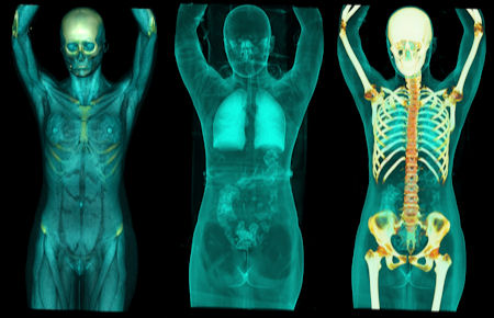
3D-rendered images using the OsiriX image fusion software. All images courtesy of Dr. Osman Ratib.
The system will also display the fused images as PET/CT over MR, PET over CT, CT over MR, and so on. The capabilities of the software can be viewed online at www.osirix-viewer.com.
The combination of PET and MR is particularly useful in radiotherapy planning, especially when treating tumors of the brain and prostate, and in some liver tumors that are treated with radiofrequency ablation. The software has also been tested to view bone metastases and in gated cardiac studies.
"PET shows the extent of metabolically active tissue in tumors, while MR shows the tissue alterations and the spatial distribution of surrounding organs. Unfortunately, very often MR does not show the exact extent of tumors due to surrounding inflammatory reactions and edema. Therefore, image fusion of PET and MR is expected to provide significant information that is not obvious from the two modalities presented separately; this can increase the accuracy and therefore confidence in diagnosis," Ratib added.
While several manufacturers are developing PET/MR hybrid scanners, with some showing an early interest in neurological applications, the technology is still largely at the prototype stage.
"The tools that are needed for physicians to deal with all these new imaging technologies are lagging behind and are still very expensive," Ratib said. "OsiriX is here to fill that gap, offering a common platform for radiologists and referring physicians to access complex data and communicate the results to each other."
The researchers expect the need for multimodality images to increase in the future. The OsiriX image fusion software is already being used in tumor boards, surgical planning sessions, and multidisciplinary clinical forums where physicians rely on image analysis to make clinical decisions.
"A three-way registration tool can provide clinicians and nonradiologists with a convenient way to review and localize focal abnormalities. That they can now interact and 'navigate' through these image sets rather than just rely on static prints provided by radiologists is a real improvement in clinical practice," Ratib concluded.
The software was designed to be fast and straightforward enough for use by radiologists as an alternative to their 3D processing workstations, as well as by clinicians who lack expertise in image processing and manipulation.
By Rob Skelding
AuntMinnie.com contributing writer
April 25, 2008
Related Reading
Cedars-Sinai explores PET's future in nuclear cardiology, April 11, 2008
Advanced visualization's benefits come with integration challenges, April 7, 2008
Integrated PET/MRI scanner developed, March 25, 2008
Fused MRI and PET improve specificity of breast MRI, August 17, 2007
Cardiac SPECT/CT fusion captures SNM's Image of the Year, June 6, 2006
Copyright © 2008 AuntMinnie.com